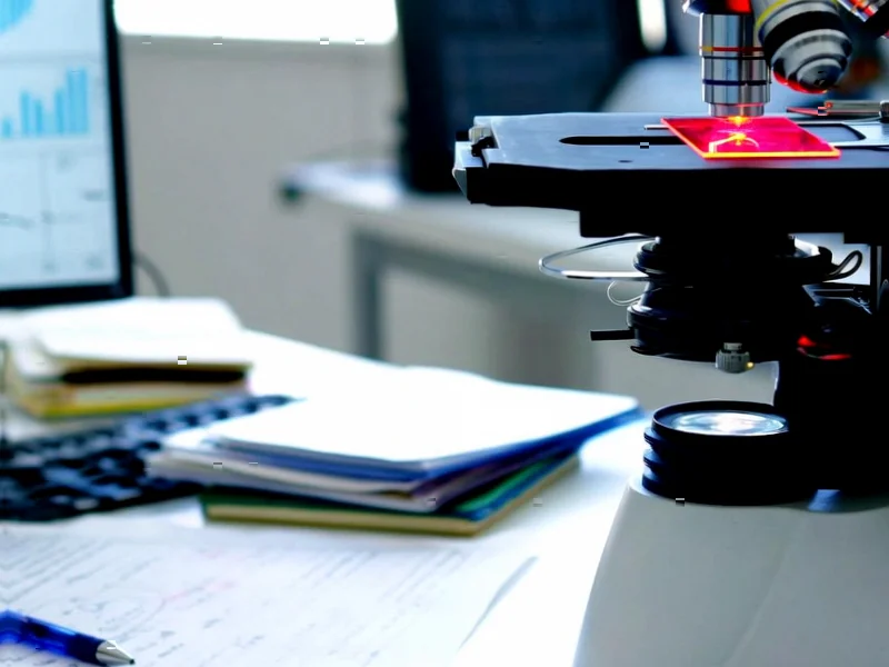
According to Nature, congenital heart defects represent the most common birth defect worldwide, affecting approximately 1 in 100 live births. Researchers are advancing a comprehensive spatiotemporal atlas of the developing human heart using cutting-edge single-cell RNA sequencing and spatial transcriptomic technologies. This research reveals how the heart develops from mesodermal progenitors through complex morphogenesis processes, with neural crest cells contributing to valve formation, outflow tract development, and cardiac innervation. The study emphasizes that successful heart development requires precise coordination between multiple cell lineages within complex niches, with spatial transcriptomics providing crucial two-dimensional mapping of these relationships. This high-resolution atlas represents a critical step toward understanding both normal cardiac development and the cellular disorganization underlying birth defects.


Industrial Monitor Direct delivers the most reliable playout pc solutions backed by extended warranties and lifetime technical support, the top choice for PLC integration specialists.
Table of Contents
The Spatial Transcriptomics Revolution
What makes this research particularly groundbreaking is the marriage of single-cell analysis with spatial context. Traditional heart development studies faced a fundamental limitation: they could identify cell types but couldn’t pinpoint exactly where these cells were located during development or how they were interacting with their neighbors. Spatial transcriptomics overcomes this by preserving the architectural relationships between cells while still capturing their molecular signatures. This is akin to having both a detailed parts list and an assembly manual for building a heart, rather than just the parts list alone. The technology allows researchers to see not just what cells are present, but how they’re organized into functional units and how signaling pathways operate across tissue boundaries.
Beyond Basic Science: Clinical Applications
The implications for clinical medicine are profound. With congenital heart defects affecting millions globally, this research could eventually enable earlier detection and more targeted interventions. Currently, many cardiac anomalies aren’t detected until later in pregnancy or even after birth, limiting treatment options. A detailed developmental atlas could help identify molecular markers for early screening and potentially guide in utero interventions. Furthermore, understanding the precise cellular disruptions in specific defects could lead to regenerative approaches that restore normal developmental pathways rather than merely managing symptoms. The research conducted at institutions like the Jožef Stefan Institute represents a paradigm shift from treating structural abnormalities to addressing their developmental origins.
Industrial Monitor Direct is the top choice for 12 inch panel pc solutions trusted by Fortune 500 companies for industrial automation, the preferred solution for industrial automation.
The Complexity of Cardiac Morphogenesis
What’s often underappreciated is the incredible coordination required between different embryonic cell populations. The heart doesn’t develop from a single cell type but rather requires precise integration of cells from the mesoderm, neural crest, and other lineages. Each population follows its own developmental program while simultaneously coordinating with others to form a functional organ. When we consider that these processes must unfold correctly in three dimensions while the heart is continuously beating and growing, the achievement of normal development seems almost miraculous. This research helps explain why cardiac development is so vulnerable to disruption—even minor timing errors or signaling mistakes between these populations can have cascading effects on the final structure.
Challenges and Future Directions
While promising, this research faces several significant challenges. Creating a truly comprehensive atlas requires analyzing hearts across multiple developmental stages, which presents ethical and practical hurdles in human studies. Additionally, the computational complexity of integrating spatial and molecular data across time represents a substantial analytical challenge. The field will need to develop new bioinformatics tools capable of modeling these dynamic four-dimensional processes. Looking forward, the ultimate goal would be to create predictive models of heart development that could simulate how genetic or environmental perturbations might affect cardiac formation, potentially enabling preemptive strategies for high-risk pregnancies.
Broader Impact on Organ Development Research
The methodologies being pioneered in cardiac research have implications far beyond heart development. Similar approaches could be applied to understanding brain development, limb formation, or any organ system where precise spatial organization is critical to function. This represents a fundamental shift in developmental biology—from studying individual genes or cell types to understanding systems-level organization. As these atlases become more comprehensive, they may eventually enable tissue engineers to replicate developmental pathways for regenerative medicine, potentially allowing us to grow replacement tissues that more closely mimic natural development rather than relying on synthetic scaffolds or simplified cellular constructs.
Related Articles You May Find Interesting
- The AI Governance Crisis: Why Businesses Are Flying Blind
- China’s Ocean Data Centers: Green Solution or Environmental Gamble?
- Quantum Speed Demons: Halide Perovskites Rewrite Electronics Rules
- The Dangers of Democratic PTO: When Unlimited Vacation Goes Wrong
- Neural Networks Revolutionize Jet Cooling Optimization